Բրենների ուռուցքները ձվարանների մակերեսային էպիթելային-ստրոմալ հազվադեպ ենթատեսակ են: Մեծամասնությունը բարորակ են, բայց որոշները կարող են չարորակ լինել[1]:
Դրանք առավել հաճախ պատահաբար հայտնաբերվում են կոնքի հետազոտության կամ լապարոտոմիայի ժամանակ[2]: Բրենների ուռուցքները շատ հազվադեպ կարող են առաջանալ այլ օրգաններում՝ ներառյալ ամորձիներում[3]:
Ներկայացում
խմբագրելՊաթոլոգիական հետազոտության ժամանակ դրանք լինում են պինդ, կտրուկ շրջագծված և գունատ դեղնավուն գույնի: 90%-ը միակողմանի է (առաջանում է մի ձվաբջջում, մյուսը՝ անփոփոխ): Ուռուցքների չափերը կարող են տարբեր լինել՝ 1 սանտիմետրից պակաս (0,39 դյույմ) մինչև 30 սանտիմետր (12 դյույմ): Չարորակ Բրենների ուռուցքները հնարավոր են, բայց հազվադեպ են հանդիպում:
Ախտորոշում
խմբագրել

Հյուսվածքաբանորեն հայտնաբերվում են անցումային էպիթելային (ուրոթելիային) բջիջներ, որոնք երկայնական ակոսներով բաժանվում են միմիանցից(սուրճի հատիկների միջուկներ), դրանք տեղակայված են առատ թելքավոր ստրոմայում։
Սուրճի հատիկների միջուկները միջուկային ակոսներ են, որոնք բացառապես պաթոգնոմիկ են սեռական լարը ստրոմալ ուռուցքի, ձվարանների հատիկավոր բջիջների ուռուցքի համար՝ հեղուկով լցված Call-Exner մարմիններով՝ հատիկավոր բջիջների միջև:
Histologically, there are nests of transitional epithelial (urothelial) cells with longitudinal nuclear grooves (coffee bean nuclei) lying in abundant fibrous stroma.
The coffee bean nuclei are the nuclear grooves exceptionally pathognomonic to the sex cord stromal tumour, the ovarian granulosa cell tumour, with the fluid-filled spaces Call–Exner bodies between the granulosa cells.[4][5]
Նմանատիպ հիվանդություններ
խմբագրելԱնցումային բջջային քաղցկեղը նույնիսկ ավելի հազվադեպ էություն է, որտեղ միզապարկի անցումային բջջային քաղցկեղի նման նորագոյացական անցումային էպիթելի բջիջները նկատվում են ձվարանների մեջ՝ առանց Բրենների ուռուցքի բնորոշ ստրոմալ/էպիթելային օրինաչափության:
Transitional cell carcinoma is an even rarer entity, in which neoplastic transitional epithelial cells similar to transitional cell carcinoma of the bladder are seen in the ovary, without the characteristic stromal/epithelial pattern of a Brenner tumour.
Հյուսվածքաբանորեն, ամորձիների Լեյդիգի բջիջների ուռուցքները և ձվարանների ստրոմալ Լեյդիգ բջջային ուռուցքները (ձվարանների հիպերանդրոգենիզմ և վիրիլիզացիա) երկուսն էլ ունեն Ռեյնկեի բնորոշ բյուրեղներ: Նույն բյուրեղները նշվել են նաև Բրենների ուռուցքների բարձր հզորության տեսադաշտում:
Histologically, Leydig cell tumours of the testes and ovarian stromal Leydig cell tumours (ovarian hyperandrogenism and virilization) both have characteristic Reinke crystals. The same crystals were also noted under high-power view in Brenner tumours.[6]
Էպոնիմ
խմբագրելԱյն անվանվել է ի պատիվ գերմանացի վիրաբույժ Ֆրից Բրենների (1877–1969), որը բնութագրել է այն 1907 թվականին։ «Բրենների ուռուցք» տերմինն առաջին անգամ օգտագործվել է Ռոբերտ Մեյերի կողմից 1932 թվականին։
It is named for Fritz Brenner (1877–1969), a German surgeon who characterized it in 1907.[7] The term "Brenner tumour" was first used by Robert Meyer, in 1932.[8]
Լրացուցիչ պատկերներ
խմբագրել-
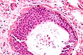 Ուոլթարդի բջիջների բույնի միկրոգրաֆիա, որից ենթադրվում է, որ առաջանում են Բրենների ուռուցքները: H&E բիծ.

Ծանոթագրություններ
խմբագրել- ↑ Marwah N, Mathur SK, Marwah S, Singh S, Karwasra RK, Arora B (April 2005). «Malignant Brenner tumour--a case report». Indian Journal of Pathology & Microbiology. 48 (2): 251–252. PMID 16758686.
- ↑ Green GE, Mortele KJ, Glickman JN, Benson CB (October 2006). «Brenner tumors of the ovary: sonographic and computed tomographic imaging features». Journal of Ultrasound in Medicine. 25 (10): 1245–51, quiz 1252–4. doi:10.7863/jum.2006.25.10.1245. PMID 16998096.
- ↑ Caccamo D, Socias M, Truchet C (May 1991). «Malignant Brenner tumor of the testis and epididymis». Archives of Pathology & Laboratory Medicine. 115 (5): 524–527. PMID 2021324.
- ↑ «Pathology Thread». University of Virginia Medical School. Արխիվացված է օրիգինալից 4 February 2006-ին.
- ↑ Ahr A, Arnold G, Göhring UJ, Costa S, Scharl A, Gauwerky JF (July 1997). «Cytology of ascitic fluid in a patient with metastasizing malignant Brenner tumor of the ovary. A case report». Acta Cytologica. 41 (4 Suppl): 1299–1304. doi:10.1159/000333524. PMID 9990262.
- ↑ Kuno Y, Baba T, Kuroda T, Teramoto M, Hirokawa N, Endo T, Saito T (October 2018). «Rare case of occult testosterone-producing ovarian tumor that was diagnosed by selective venous hormone sampling». Reproductive Medicine and Biology. 17 (4): 504–508. doi:10.1002/rmb2.12213. PMC 6194242. PMID 30377407.
- ↑ Lamping JD, Blythe JG (September 1977). «Bilateral Brenner tumors: a case report and review of the literature». Human Pathology. 8 (5): 583–585. doi:10.1016/S0046-8177(77)80117-2. PMID 903146.
- ↑ Philipp EE, O'Dowd MJ (2000). The history of obstetrics and gynaecology. Carnforth, Lancs: Parthenon. էջ 586. ISBN 978-1-85070-040-1.
External links
խմբագրել| Դասակարգում |
|---|
- «Brenner tumour». Medcyclopaedia. GE. Արխիվացված է օրիգինալից 2012-02-05-ին.
- Histology at University of Utah
Կաղապար:Soft tissue tumors and sarcomas Կաղապար:Breast cancer/urogenital neoplasia